Diameter reduction of the proximal long saphenous vein after ablation of a distal incompetent tributary
Diameter reduction of the proximal long saphenous vein after ablation of a distal incompetent tributary
SUMMARY
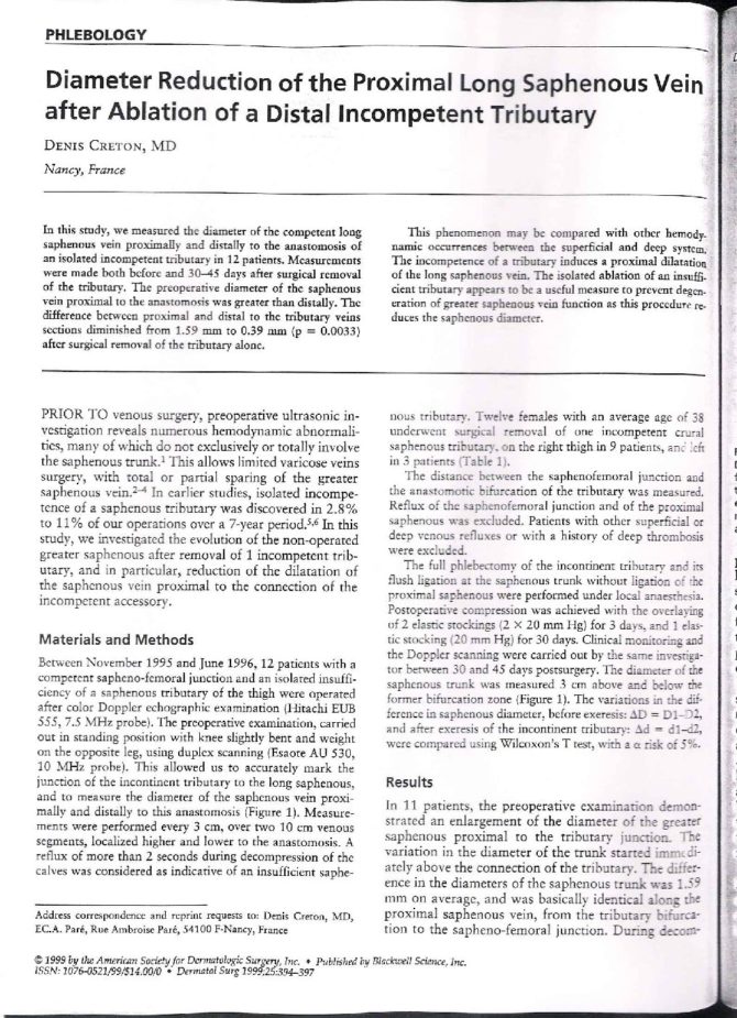
In this study we measured the diameter of the competent long saphenous vein proximally and distally to the anastomosis of an isolated incompetent tributary in 12 patients. Measurements were made both before and 30-45 days after surgical removal of the tributary. The preoperative diameter of the saphenous vein proximal to the anastomosis was greater than distally. The difference between proximal and distal to the tributary veins sections diminished from 1.59 mm to 0.39 mm (p=0.0033) after surgical removal of the tributary alone.
This phenomenon may be compared with other hemodynamic occurrences between the superficial and deep system. The incompetence of a tributary induces a proximal dilatation of the long saphenous vein. The isolated ablation of an insufficient tributary appears to be a useful measure to prevent degeneration of greater saphenous vein function as this procedure reduces the saphenous diameter.
INTRODUCTION
Prior to venous surgery, preoperative ultrasonic investigation reveals numerous hemodynamic abnormalities many of which do not exclusively or totally involve the saphenous trunk (1). This allows limited varicose veins surgery, with total or partial sparing of the greater saphenous vein (2-4). In earlier studies, isolated incompetence of a saphenous tributary was discovered in 2.8% to 11% of our operations over a 7-year period (5,6). In this study, we investigated the evolution of the non-operated greater saphenous after removal of one incompetent tributary, and in particular, reduction of the dilatation of the saphenous vein proximal to the connection of the incompetent accessory.
MATERIALS AND METHODS
Between November 1995 and June 1996, 12 patients with a competent sapheno-femoral junction and an isolated insufficiency of a saphenous tributary of the thigh were operated after color Doppler echographic examination (Hitachi EUB 555, 7.5 MHz probe). The preoperative examination, carried out in standing position with knee slightly bent and weight on the opposite leg using duplex scanning (Esaote AU 530, 10 MHz probe). This allowed us to accurately mark the junction of the incontinent tributary to the long saphenous, and to measure the diameter of the saphenous vein proximally and distally to this anastomosis (figure I). Measurements were performed every 3 cm, over two 10 cm venous segments, localized higher and lower to the anastomosis. A reflux of more than 2 seconds during decompression of the calves was considered as indicative of an insufficient saphenous tributary. Twelve females with an average age of 38 underwent surgical removal of one incompetent crural saphenous tributary, on the right thigh in 9 patients, and left in 3 patients (table I).
The distance between the sapheno-femoral junction and the anastomotic bifircation of the tributary was measured. Reflux of the sapheno-femoral junction and of the proximal saphenous was excluded. Patients with other superficial or deep venous refluxes or with a history of deep thrombosis were excluded.
The full phlebectomy of the incontinent tributary and its flush ligation at the saphenous trunk without ligation of the proximal great saphenous vein were performed under local anaesthesia. Postoperative compression was achieved with the overlaying of 2 elastic stockings (2 X 20 mm Hg) for 3 days, and 1 elastic stocking (20 mm Hg) for 30 days. Clinical monitoring and the Doppler scanning were carried out by the same investigator between 30 and 45 days post-surgery. The diameter of the saphenous trunk was measured 3 cm above and below the former bifurcation zone (Figure 1). The variations in the difference in saphenous diameter, before exeresis : *D=D1-D2, and after exeresis of the incontinent tributary : *d=d1-d2, were compared using Wilcoxon’s T test, with a * risk of 5%.
RESULTS
In eleven patients, the preoperative examination demonstrated an enlargement of the diameter of the greater saphenous proximal to the tributary junction. The variation in the diameter of the trunk started immediately above the connection of the tributary. The difference in the diameters of the saphenous trunk was 1.59 mm on average, and was basically identical along the proximal saphenous vein, from the tributary bifurcation to the sapheno-femoral junction. During decompression of the incontinent tributary, a slow reflux, lasting more than one second, was recorded on three occasions (one case during the first examination, and in two other cases during the second examination). This reflux was located on the proximal saphenous trunk, in the area next to the bifurcation; there was no incompetence during compression-decompression of the calf or during Valsalva’s maneuver.
Following operation, no incompetence was observed in the saphenous trunk. In 7 cases the proximal saphenous trunk had the same diameter as the distal saphenous trunk, with no difference in diameter at the level of the former bifurcation (Table I). In the 5 remaining cases, the difference in diameter persisted, but was far less than prior to the operation. The average difference in diameter above and below the former bifurcation was 0.39 mm. Statistical comparison of the reductions in diameter of the saphenous trunk before and after the operation shows that they were significantly different (Wilcoxon’s T test p = 0.0033).
Nine patients were seen one year after the operation. Eight of these patients had no saphenous deformation, and the diameter of the saphenous trunk was homogenous and unchanged. One patient had another isolated incompetence of a saphenous tributary located several centimetres above the previous one. It had the same deformation of the saphenous trunk with the proximal section greater in diameter than the distal.
Figure 1
Comparison between the modifications in diameter Delta D=D1-D2 et Delta d=d1-d2 before and after the exeresis of the insufficient tributary. D1 and d1 are the diameters of the saphenous trunk located above the insufficient tributary before and after the exeresis of the tributary. D2 and d2 are the diameter of the saphenous trunk located below the incompetent tributary before and after the exeresis of the tributary.
Table I : Distribution of the 12 Cases Before and After Exeresis of the Insufficient Tributary.
| Diameter of the saphnenous trunk (mm) | ||||||||
|---|---|---|---|---|---|---|---|---|
| Before exeresis | After exeresis | |||||||
| age | side | D1 | D2 | Delta D | d1 | d2 | Delta d | |
| 35 | D | 4,8 | 3,5 | 1,3 | 3,5 | 3,4 | 0,1 | |
| 40 | D | 4,5 | 3,5 | 1 | 3,7 | 3,7 | 0 | |
| 47 | G | 4,7 | 3,2 | 1,5 | 5 | 5 | 0 | |
| 35 | D | 4,3 | 2,7 | 1,6 | 2,4 | 2,4 | 0 | |
| 48 | D | 3,8 | 2,4 | 1,4 | 3 | 2,7 | 0,3 | |
| 35 | D | 5,7 | 4,2 | 1,5 | 5,9 | 4,5 | 1,4 | |
| 29 | D | 4,5 | 4,5 | 0 | 3,5 | 3,5 | 0 | |
| 29 | D | 4 | 2,7 | 1,3 | 3,5 | 3,5 | 0 | |
| 56 | G | 4,3 | 1,3 | 3 | 2,5 | 1,5 | 1 | |
| 37 | G | 6 | 3,5 | 2,5 | 3 | 3 | 0 | |
| 35 | D | 7 | 5 | 2 | 5 | 5 | 0 | |
| 30 | D | 7 | 5 | 2 | 4 | 2,5 | 1,5 | |
D1 : proximal diameter of the saphenous trunk. D2 : distal diameter of the saphenous trunk
D2 et D1 : diameter of the saphenous trunk above and below the insufficient tributary.
d1 : diameter of the trunk above the former location of the tributary. d2 : diameter of the trunk below
d2 et d1 : diameter of the saphenous trunk above and below the former location of the tributary.
Difference in diameter of the saphenous trunk before the exeresis delta D = D1 – D2
Difference in diameter of the saphenous trunk after the exeresis delta d = d1 – d2
Delta D is greater than Delta d (difference is significant p = 0,0033)
DISCUSSION
Despite of the limited number of patients included in this study, the results are significant. They show that when there is a major dilatation of the saphenous trunk above an incontinent tributary, removal of the incontinent tributary allows the proximal saphenous trunk to regain its normal diameter.
- The dilatation of the proximal saphenous trunk seems paradoxical, and may be similar to Venturi’s phenomenon. According to Bernoulli, the reduction in flow and speed in the saphenous segment located above the anastomosis of the tributary leads to a reduction in kinetic energy and an increase in pressure. But whilst this hypothesis is theoretically legitimate, it is unlikely that it can be applied to this particular case, where circulatory speed (few centimetres per second) is slow, and subject to the intermittent nature of venous flow.
The explanation of this hemodynamic model could include the speed of reflux required for valve closure (this speed was assessed at 30 cm/s by van Bemmelen (7) for deep veins). The incompetence of the saphenous tributary induces a slow reflux, which is probably insufficient to close the valve located just above the anastomosis of the tributary. The slow and continuous reflux maintained at the level of this valve together with the normal flow of the saphenous trunk may produce a venous stasis due to the excess volume and a dilatation of the intervalvular space located above the incompetent tributary.
Furthermore, some circulatory factors linked to circulatory speed play a role regarding arterial endothelial vasomotricity (Nitric Oxide) (8, 9). Concerning varicose vein disease it has been proved that endothelin 1 is elevated with a diminution of the contractile response. It may contribute to the pathogenesis of varicose vein dilatation (10). It is possible that these endothelial factors linked to blood flow play a role in the enlargement of the saphenous trunk because of an alteration in the endothelial function.
- The reduction in diameter of the primary proximal trunk after removal of the incompetent secondary trunk is a hemodynamic model that enables us to explain some observations. This hemodynamic model is applicable to the venous bifurcations between a competent primary trunk and an incompetent secondary trunk.
It allow us to explain the dilatation of the deep venous system that accompanies reflux in the superficial venous system. This phenomenon was first described by Trendelenburg (11). Fischer (12), in a phlebographic study of 68 cases stressed that the dilatation of the deep veins was greater in patients who had no post-thrombotic sequela. This might lead us to think that this phenomenon can only have a dilatatory effect on the deep system if the latter is free of fibrous scarring. Many authors (11,12) have described the reversibility of both the dilatation and the reflux on the deep primary venous trunk, through the removal of the superficial secondary trunk. Almgren (13) has shown a significantly larger proportion of deep venous reflux in groups of patients with varices or recurrent varices compared to groups of patients who are healthy or who have undergone removal of their varices. Other authors (14) have observed a reduction in the diameter of deep veins in varicose patients who have been operated upon.
Concerning the sapheno-popliteal junction, Somjen (15) has shown that removal of the incompetent lesser saphenous vein made the reflux disappear (the reflux often being associated with the popliteal vein when the popliteal valve was competent). He explains that the extension of the reflux to the deep system is due to an increase in pressure at this level. Reflux at this level is due to both dilatation of the deep system and the aspiration of the blood located in the non-valvulated veins near to the sapheno-popliteal junction by the lesser saphenous reflux (16).
The same phenomenon was observed at the sapheno-femoral junction by Walsh (17). He demonstrated that the reflux in the superficial femoral vein disappeared in 27 out of 29 of limbs operated on for varices. In this study, reflux measurements were made on the femoral vein below the bifurcation, and not at or above the bifurcation, as was the case in Somjen’s study (15). Among 17 patients with incompetent femoral and long saphenous veins, Sales (18) showed that in 16 cases, a saphenectomy restored normal deep hemodynamics without reflux. The only patient whose deep venous incompetence was not corrected by saphenectomy was a patient with popliteal and femoral vein incompetence (grade IV Kistner).
With regard to greater saphenous incompetence associated with an incompetent tributary, it is difficult to suggest a saphenous overload mechanism. This hemodynamic model was described by Vidal-Michel (19). He showed that the removal of an incompetent saphenous tributary could reduce the diameter of the sapheno-femoral junction, and could even cause the incompetence of the sapheno-femoral junction or proximal trunk to disappear. The disappearance of the reflux sometimes located on the proximal trunk may simply be due to the reduction in the diameter of the saphenous trunk. Sclerotherapy of saphenous trunks, by reducing the diameter of the saphenous trunk, has in certain cases also allowed normal saphenous function to be restored (20).
- The origin of lower limb primary venous reflux appears to be a local or multifocal process (21). There are several types of progressive degradation of saphenous function over time but in certain cases varicose illness begins with tributary incompetence. In a group of 607 patients (6), we demonstrated that isolated incompetence of a saphenous tributary was present in a group that was 10 years younger than the group of patients with total incompetence of the saphenous trunk. Moreover in this study, one patient developed the same deformation of the trunk associated with a new refluxing tributary.
In a similar study of varicose patients of different ages, ALMGREN (22) noticed that segmented saphenous insufficiency occurred approximately 10 years before complete saphenous insufficiency. It has also been demonstrated (23) that a difference between the pathology of the saphenous trunk and the tributaries concerning the contraction response for sarafotoxin S6c. This suggests that the pathology of the tributaries is more important than that of the trunk. In the specific cases where the varicose illness appears to progress from the tributaries towards the trunk, the ablation of incompetent saphenous tributaries can be a preventive technique. This preventive technique will enable us to restrict the progressive degradation of the saphenous function, and to preserve the saphenous trunk for a longer time.
Long term studies and a larger number of patients must confirm this preliminary study. Such studies will also give us a better understanding of the general evolution of varicose disease all throughout the long saphenous system.
REFERENCES
- LARGE J. Surgical treatment of saphenous varices, with preservation of the main great saphenous trunk. J Vasc Surg 1985;2:886-891.
- GOREN G. Primary varicose veins : topographic and hemodynamic correlations. J Cardiovasc Surg 1990;31:672-677
- GOREN G. Primary varicose veins : hemodynamic principles of surgical care (The case for ambulatory stab avulsion technique).VASA 1991;20:-365-368.
- KOYANO K, SAKAGUCHI S. Selective stripping operation based on doppler ultrasonic findings for primary varicose veins of the lower extremities. Surgery 1988;103:615-619.
- CRETON D. Influence des examens ultrasonores préopératoires pour une chirurgie d’exérèse plus conservatrice. Phlébologie 1994;47:227-234.
- CRETON D. Classification des varices sur 607 échomarquages : aspect évolutif de la maladie variqueuse. in:Negus D, Jantet C, Colleridge Smith PD, eds, Phlebology 95,Suppl 1, London, Springer Verlag,1995:160-163.
- Van BEMMELEN PS, BEACH K, BEDFORD G, STRANDNESS DE. The mechanism of venous valve closure. Its relationship to the velocity of reverse flow. Arch Surg 1990;125:617-619.
- ADAMS MR, ROBINSON J, SORENSEN KE, DEANFIELD JE, CELERMAJER DS, Normal range for brachial artery flow-mediated dilatation: a non-invasive ultrasound test of arterial endothelial function. J Vasc Invest. 1996;2:3:146-150.
- LIEBERMAN EH, KNAB ST, CREAGER MA. Nitric oxide mediates the vasodilatator response to flow in humans (abstrast). Circulation 1994;90(suppl):1-138.
- LOWEL RC, GLOVICZKI P, MILLER VM. In vitro evaluation of endothelial and smooth muscle function of primary varicose veins. J Vasc Surg 1992;16:679-686.
- TRENDELENBURG F. Ueber die Unterbindung der Vena saphena magna bei Unterschenkelvaricen. Beitr Z Klin Chir 1891;7:195-210.
- FISCHER H, SIEBRECHT H. Das Kaliber der tiefen Unterschenkelvenen bei der primaren Varicose und beim postthrombotischen Syndrom (Eine phlebographische Studie). Der Hautarzt 1970;21:205-211.
- ALMGREN Bo, ERIKSSON I. Primary deep venous incompetence in limbs with varicose veins. Acta Chir Scand 1989;155:455-460.
- STRANZENBACH W, HACH W. Phlebographische Verlaufsbeobachtungen der sekundaren Popliteal und Femoralveneinsuffizienz bei Stammvarikose. Phlebol 1991;20:25-29.
- SOMJEN GM, ROYLE JP, FELL G, ROBERTS AK, HOARE MC, TONG Y.Venous reflux patterns in the popliteal fossa. J Cardiovasc Surg 1992;33:85-91.
- BROWSE NL, BURNAND KG, LEA THOMAS M, Primary (non-thrombotic) deep vein incompetence. in BROWSE NL, BURNAND KG, LEA THOMAS M, (eds). Diseases of the veins, pathology, diagnosis and treatment. London, Edward Arnold. 1988:253-268.
- WALSH JC, BERGAN JJ, BEEMAN S, COMER TP. Femoral venous reflux abolished by greater saphenous vein stripping. Ann Vasc Surg 1994;8:566-570.
- SALES CM, BILOF ML, PETRILLO KA, LUKA NL, Correction of lower extremity deep venous incompetence by ablation of superficial venous reflux. Ann Vasc Surg 1996;10:186-189.
- VIDAL-MICHEL JP, BOURREL Y, EMSALLEM J, BONERANDI JJ. Respect chirurgical des crosses saphènes internes modérément incontinentes par effet siphon chez les patients variqueux. Phlébologie 1993;46:143-147.
- SCHADECK M. Le reflux sur valvules saines en grande saphène. Phlébologie 1991;44:603-613.
- LABROPOULOS N, GIANNOUKAS AD, DELIS K, MANSOUR MA, KANG SS, NICOLAIDES AN, LUMLEY J, BAKER WH. Where does venous reflux start ? J Vasc Surg 1997;26:736-742.
- ALMGREN Bo, ERIKSSON I. Valvular incompetence in superficial, deep and perforator veins of limbs with varicose veins. Acta Chir Scand 1990;156:69-74.
- BARBER DA, WANG XW, GLOVICZSKI P, MILLER VM. Characterization of endothelin receptors in human varicose veins. J Vasc Surg. 1997;26:61-69.
| Date | 1999 |
| Awards | Dermatol Surg 1999;25:1-5. Dr Denis CRETON EC. A. PARE, Rue Ambroise Paré, 54100 F-NANCY Tel : +33 (0)3 83 95 54 00 Fax : +33 (0)3 83 95 54 23 |