Recurrence of popliteal varicose veins after SSV excision
Recurrence of popliteal varicose veins after SSV excision
Recurrence of popliteal varicose veins has long been attributed to insufficient excision of an incompetent short saphenous vein. Indeed, recurrence is still frequent after surgery of the short saphenous vein. In order to assess the risk of insufficient excision and study the mechanism of recurrence, we reviewed 125 popliteal procedures for recurrence after excision of an incompetent short saphenous vein.
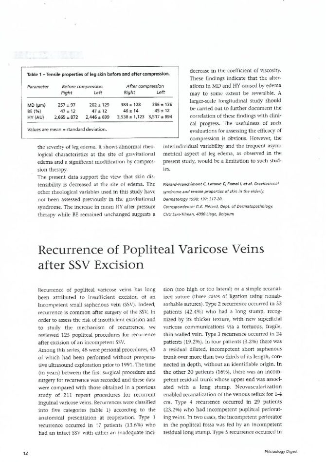
Among this series, 48 were personal procedures, 43 of which had been performed without preoperative ultrasound explorations prior to 1991. The times (in years) between the first surgical procedure and the one for recurrence were recorded and compared to those of a previous study on 211 repeat procedures for recurrent inguinal varicose veins [1]. Recurrences were classified into five categories (table I) according to the anatomic presentation at reoperation.
- Type 1: 17 patients had an intact short saphenous (13.6%) with either an inadequate incision ( too high or too lateral) or a simple recanalized suture (3 cases of ligation using non-adsorbable sutures).
- Type 2: 53 patients had a long stump recognized by its thicker texture (42.4%) with new superficial varicose communications via an tortuous fragile, thin walled vein.
- Type 3: 4 patients had a residual dilated incompetent short saphenous trunk over more than 2/3 of its length, connected in depth, without an identifiable origin (3.2%). Twenty patients had both an incompetent residual trunk whose upper end was associated with a long stump. The neovascularization enabled recanalization of the venous reflux along 1 to 4 cm (16 %).
- Type 4: 29 patients had incompetent popliteal perforating veins. In 2 cases, the incompetent perforator in the popliteal fossa was fed by an incompetent residual long stump (23.2%).
- Type 5: 2 patients developed recurrence on a new varicose communication which followed the posterior nerves of the thigh. These 2 patients had undergone surgery for an incompetent short saphenous vein joining in the first case the sciatique nerve and in the second case the peroneal saphenous nerve. The resection had been performed near the nerve. All the junctions of residual stumps were located in the popliteal fossa between 1 and 8 cm exept for one located 17 cm above the popliteal fold.
Among our personal series, 43 reoperations for recurrence were performed without preoperative duplex Doppler exploration. Among them, excision was insufficient in 38 (table II). The 5 patients reoperated after preoperative ultrasound exploration had a popliteal perforating vein. Recurrence with a popliteal perforator was significantly more frequent in men than in women (table III). The time of reoperation for popliteal recurrence after surgery of the short saphenous ( 50% at 6 years) was significantly shorter than the time to reoperation for inguinal recurrence after surgery of the greater saphenous (50% at 12 years). Insufficient excision was observed in 75.2% of the cases, but 23.2% of the recurrences were due to the development of an incompetent popliteal perforating veins.
These perforating veins were residual stumps of the short saphenous with complex pathways, unrecognized perforating veins associated with the short saphenous at the first operation or a new developing in several perforating vessels described in the popliteal fossa. The lack of preoperative ultrasound data made it difficult to interpret these recurrences. The development of perforating veins in the popliteal fossa is a type of recurrence which is probably the expression of particular hemodynamic phenomena in the popliteal venous circulation. These phenomena probably involve the flexion of the popliteal vein, the contraction of the calf muscles, and also popliteal valvular incompetence frequently demonstrated in patients who develop short saphenous vein insufficiency.
TABLE I
Anatomic types of popliteal recurrences. Anatomic types 1, 2 and 3 result from insufficient excision of an incompetent small saphenous (75%), type 4 from the development of perforating veins in the popliteal fossa, and type 5 from varicose communications with the vasa-nervorum of the posterior nerves of the thigh.
| Type | Anatomy of recurrence | N=125 | % | |
| 1 | Intact short saphenous | 17 | 13.6 | |
| 2 | Long stump | 53 | 42.4 | |
| 3 | Saphenous trunk Long stump + saphenous trunk |
4 20 |
3.2 16 |
75% |
| 4 | Perforating vein in popliteal fossa | 29 | 23.2 | |
| 5 | Sciatic vein | 2 | 1.6 |
TABLE II
Anatomic types of recurrence by preoperative duplex-Doppler results (48 personal cases).
| Without Doppler ultrasound | With Doppler ultrasound | |
| Insufficient excision | 38 | 0 |
| Vein in popliteal space | 5 | 5 |
TABLE III
Popliteal recurrence repair by sex (n=125). The risk of developing a popliteal perforating vein type recurrence after excision of an incompetent short saphenous is significantly higher in men than in women (p<0.05).
| Men | Women | |
| Recurrent popliteal varicose veins | 23.2% | 76.8% |
| Recurrent popliteal varicose veins by a perforating vein in the popliteal space | 41.4% | 58.6% |
REFERENCES
- CRETON D. hypotheses on the aetiology of recurrences of varices of the great saphenous system : anatomical study of 211 patients. in: Negus D, Jantet G, Colleridge-Smith PD, (eds), Phlebology 95, Suppl. 1, London, Springer-Verlag, 1995: 164-168.
| Date | 2000 |
| Awards | Phlebology Digest 2000;13:12-3 D. CRETON EC. AP, rue A. PARE, 54100 F-NANCY. Tel : +33 (0)3 83 95 54 00, Fax : +33 (0)3 83 95 54 23 |