Prosthetic material interposition on the crossectomy stump in varicose vein recurrence surgery: preliminary report on the prevention of angiogenesis
Prosthetic material interposition on the crossectomy stump in varicose vein recurrence surgery: preliminary report on the prevention of angiogenesis
SUMMARY
149 consecutive patients presenting a groin varicose veins recurrence underwent a re-exploration of the previous crossectomy site, with implantation of a Gore-Tex e-PTFE patch on the femoral stump with a lateral approach, under loco regional anesthesia, in one day surgery. The aim was to curb the risk of new recurrence. All the patients were re-examined clinically and with ultrasound doppler 45 days after the operation. Apart from 4 infection complications and 2 delays in scarring, the patch was always well tolerated and was never been rejected. 39 patients were re-examined after 5 years and 4 patients only presented a new varicose veins recurrence in the groin, but these varicose veins were fed by another vein than the femoral vein. No new varicose veins were connected with the femoral vein. This preliminary report on 39 patients only, the good tolerability of the ePTFE patch and the low morbidity associated with its implantation, made us recommend isolation of the crossectomy stump as a means of preventing a new groin surgical re-exploration for a new recurrence.
INTRODUCTION
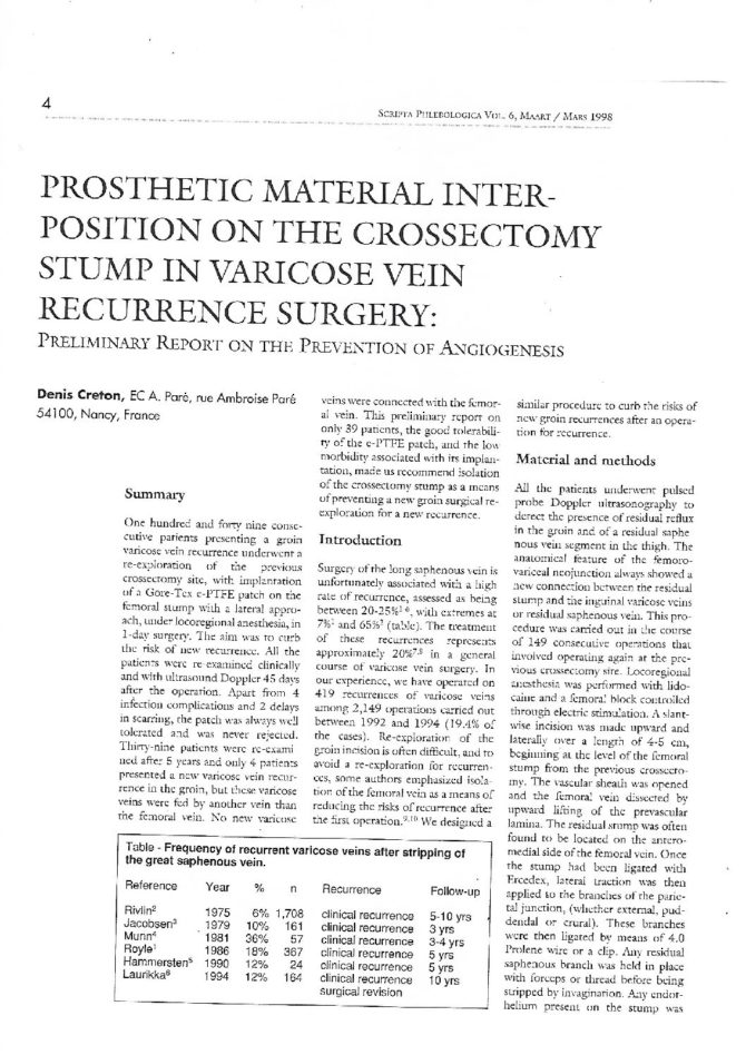
Surgery of the long saphenous vein is unfortunately associated with a high rate of recurrence assessed as being between 20% and 25% [1-6], with extremes at 7% [1] and 65% [2] (Table I).The treatment of these recurrences represents approximately 20% [7,8] in a general course of varicose vein surgery. In our experience we have operated on 419 recurrences of varicose veins among 2,149 operations carried out between 1992 and 1994 (19,4% of the cases). Re-exploration of the groin incision is often difficult, and to avoid a re-exploration for recurrences some authors emphasized isolation of the femoral vein as a means of reducing the risks of recurrence after the first operation [9,10]. We designed a similar procedure to curb the risks of new groin recurrences after an operation for recurrence.
Tableau 1 : Frequency of recurrent varicose veins after stripping of the great saphenous vein
| Reference | year | % (n) | recurrence | followup |
| RIVLIN [2] | 1975 | 6% 1708 | clinical recurrence | 5-10 yrs |
| JACOBSEN [3] | 1979 | 10% 161 | clinical recurrence | 3 yrs |
| MUNN [4] | 1981 | 36% 57 | clinical recurrence | 3-4 yrs |
| ROYLE [1] | 1986 | 18% 367 | clinical recurrence | 5 yrs |
| HAMMERSTEN [5] | 1990 | 12% 24 | clinical recurrence | 5 yrs |
| LAURIKKA [6] | 1994 | 12% 164 | clinical recurrence surgical revision | 10 yrs |
MATERIAL AND METHOD
All the patients underwent a pulsedprobe Doppler Ultrasonography to detect the presence of a residual reflux in the groin and of a residual saphenous vein segment in the thigh. The anatomical feature of the femoro-variceal neojunction always showed a new connection between the residual stump and the inguinal varicose veins or residual saphenous vein. This procedure was carried out in the course of 149 consecutive operations that involved operating again at the previous crossectomy site. Locoregional anesthesia was performed with lidocaine and a femoral block controlled through electrical stimulation. A slantwise incision was made upward and laterally over a length of 4 to 5 cm, beginning at the level of the femoral stump from the previous crossectomy. The vascular sheath was opened and the femoral vein dissected by upward lifting of the prevascular lamina. The residual stump was often found to be located on the anteromedial side of the femoral vein. Once the stump had been ligated with Ercedex, lateral traction was then applied to the branches of the parietal junction, (whether external, pudental or crual). These branches were then ligated by means of 4.0 Prolene wire or a clip. Any residual saphenous branch was held in place with forceps or thread before being stripped by invagination. Any endothelium present on the stump was micro-coagulated and stitches of 4.0 Prolene wrapped around the stump.The obliteration of the residual stump had been effected in two ways.
- By lateral femoral clamping and complete resection of the stump followed by a continuous 6-0 Prolene wire tranverse suture of the femoral vein for the 10 first patients.
- By putting a transverse suture (whithout opening the femoral vein) for burying the stump into the venous adventicia in order to prevent any connection from forming between the endothelial cells of the stump’s lumen and subcutaneous tissues for the 139 following patients.
In each case, by placing an e-PTFE membrane, 1 centimeter thick, on the stump, a barrier was erected between the femoral stump and the subcutaneous tissues (Figure 1). This patch was trimmed to about 2 to 3 cm2, approximately 2 cm in length and 1 cm in width, placed over the femoral stump so as to greatly overlap the suture site (Figure 2).Two groups of patients were separated. The first group concerned 40 patients in which the patch was anchored over the femoral vein by means of two Prolene 5/0 stitches, with the same prolene used for the suture, or by means of another thread fixed on the transverse suture (Figure 1).The patch was also glued in place with biological glue.The second group concerned 109 patients ; in these cases no thread was used for the fixation of the patch. In order to prevent the patch from moving, the dissection was made as small as possible. We tried to carry out a cavity of dissection with a dimension corresponding to that of the patch to avoid a displacement of the patch. In each case the patch was glued in place with biological glue applyied over and under the patch. The incision was then closed without drainage by means of a slow-resorbing intradermal suture. All the patients had a duplex ultrasound control and a clinical examination 45 days after the operation. Among these, only 39 patients have been re-examined after 5 years long term follow up. A new recurrence was only searched on the medial aspect of the leg along the long saphenous vein area.
Fig 1: Fixation of the patch over the suture
Fig 2: The patch is fixed over the femoral vein by means of 2 Prolene 5/0 stitches

RESULTS
The operation, performed on an outpatient basis, involved 134 patients ; 15 other patients were hospitalized, either for personal reasons or because of an adverse social environment or medical problems such as : postoperative asthenia, too long a period of anesthesia, or intercurrent disorders mainly affecting the nose and throat.
Short term results :
At the beginning of our experiment, twice during the operation, a lateral tear in the femoral vein, caused by disinsertion of one branch prevented us from applying the patch. One of the patients, who was obese, complained of chest pain a few hours after the operation. He was hospitalized for cardiopulmonary observation which did not reveal the presence of any thromboembolic complications. In 2 cases, there was a delay in scarring (2 weeks). Infectious complications were recorded in 4 other cases (one abscess had to be drained under general anesthesia at day 8 postoperatively, but did not require removal of the patch). One case presented with an hematoma which was spontaneously evacuated without infectious complications. In all cases tolerability of the patch was high and no rejection occurred. No lymphorrhea was observed postoperatively. Doppler ultrasound examination did not reveal any collection of fluid nor any deformation of the venous femoral wall except in 10 cases at the beginning of our experience. In these cases the purse-string ligature for burying the stump had been responsible for a small and located deformation of the anterior aspect of the femoral vein.
Long term results :
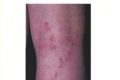
Long term follow up was studied only in 39 patients. Among these, 4 patients presented varicose vein in the calf, with a small varicious network in the thigh which could be treated by sclerotherapy of phlebectomy. Four other patients presented varicose veins in the upper part of the thigh with a diametre of more than 4 mm.The Ultrasound doppler examination of these veins near the groin area showed that theyre were not connected with the femoral vein. Although the new varicose veins were very close to the femoral vein, they were fed by abdominal subcutaneous veins or puddental veins. In each case, the patch was clearly seen and sometimes the patch was located just between the femoral vein and the nearer new varicose veins (Figure 3). Once the new varicose vein passed through a lymphatic node (Figure 4). 31 patients had no varicose vein recurrences and had a good cosmetic results.
Fig 3 : Varicose veins recurrence without connection with the femoral vein
Fig 4 : New varices passing through a lymphatic node (angiogenesis)
DISCUSSION
Study of the various anatomical features associated with recurrent varicose veins showed this condition to be caused by two mechanisms [11].
- Mechanical recurrences in which residual hemodynamic hyperpressure appears to be a key factor in explaining recurrence is actually due to mistakes. Hemodynamical recurrences are generally induced by sugical mistakes : partial resection of the saphenofemoral junction, persistance of a femoral vein tributary.
- Histological recurrences, e.g., diffuse varicosities without residual reflux, in which the recurrence is attributable to angiogenetic biological phenomena [12,13].
It follows that in order to prevent recurrence following complete crossectomy, potential connections between the femoral vein and the superficiel subcutaneous tissues should be avoided [14-18]. Prefemoral isolation has been suggested for first-line crossectomy by suturing the cribiform fascia in front of the femoral vein [9]. Implanting prosthetic material in front of the vein has also been suggested for the surgical cure of short saphenous vein reflux [10]. Since the fascia cannot be sutured in front of the vein in revision surgery following recurrences, we elected to use a sheet of Gore-Tex sheet surgical membrane. The low porosity of the e-PTFE patch (under one µ ), which is used for pericardial [19] or peritoneal [20] reconstruction, seemed worthy of consideration to prevent any cellular ingrowth. In this study angiogenesis appeared to be involved in the 4 important new recurrences in the thigh according to the ultrasound feature of the varicose vein which passed through lymphatic nodes. Actually the 4 new groin recurrences did not concern a re-neosaphenofemoral junction. In that case re-exploration of the previous crossectomy area is not necessary. The treatment can be performed by scleroterapy or phlebectomy only. The low infectious risk is probably due to the minimal dissection and to the lateral approach of the femoral vein also advocated by others [21]. The cutaneous incision, placed a little further away from the patch implantation site, may also minimize the risk of postoperative infection.
CONCLUSION
Recurrences of the hemodynamic type, such as the formation of major femoro-variceal neojunctions or of femorosaphenous neojunctions on a residual saphenous vein can often be avoided by applying a rigourous surgical technique. Minor femoro-variceal neojunctions on minor backward flows (angiogenesis), however, do not depend upon the surgical technique used. In such a case the good tolerability of the e-PTFE patch and the low morbidity associated with its implantation in front of the femoral stump made us recommend isolation of the crossectomy stump as a means of preventing this type of recurrence involving the femoral vein. But comparative studies and a complete long term follow up will be necessary to assert the definitive usefullness of this technique.
REFERENCES
- ROYLE JP. Recurrent varicose veins. World J Surg 1986;10:944-953.
- RIVLIN S. The surgical cure of primary varicose veins. Br J Surg 1975;62:913-917.
- LAURIKKA J, SISTO T, SALENIUS JP, TARKKA M, AUVINEN O. Long saphenous vein stripping in the treatment of varicose veins : self-and surgeon-assessed results after ten years. Phlebology 1994;9:13-16.
- HAMMARSTEN J, PEDERSEN P, CEDERLUND CG, CAMPANELLO M. Long saphenous vein saving surgery for varicose veins. A long term follow-up. Eur J Vasc Surg 1990;4:361-364.
- JACOBSEN BH.The value of different forms of treatment for varicose veins. Br J Surg 1979;66: 182-184.
- MUNN SR, MORTON JB, MACBETH AG, MACLEISH AR. To strip or not to strip the long saphenous vein? a varicose veins trial. Br J Surg 1981;68:426-428.
- DAVIES GC. The Lothian surgical audit. Medical Audit News 1991;1:26-27.
- NEGUS D. Reccurent varicose veins : A national problem. Br J Surg 1993;80:823-824.
- GLASS GM. Neovascularization in recurrence of varices of the Great Saphenous Vein in the Groin : Surgical Anatomy and Morphology. Vasc Surg 1989;23:435-442.
- SHEPPARD M. The incidence diagnosis and management of sapheno-popliteal incompetence, Phlebology 1986;1:23-32.
- CRETON D. Hypothèses étiologiques des récidives variqueuses Saphène internes : Etude anatomique sur 211 cas. Phlebology’95, D Negus et al. (eds).Phlebology 1995. Suppl.1,164-168.
- COUFFINHAL JC, BOUCHEREAU R. Angiogénèse et insuffisance veineuse superficielle » la maladie induite ». Phébologie 1990;43:609-613.
- FORM DM. Endothelial cell proliferation during angiogenesis. Lab Invest 1980;65:1351-1361.
- GLASS GM. Neovascularization in recurrence of varices of the Great Saphenous Vein in the Groin : Phlebography. Angiology 1988;39:577-582.
- GLASS GM. Neovascularization in recurrence of varices of the Great Saphenous Vein in the Groin : Surgical Anatomy and Morphology. Vasc Surg 1989;23:435-442 .
- GLASS GM. Neovascularization in recurrence of the varicose Great Saphenous Vein following transection . Phlebology 1987;2:81-91.
- GLASS GM. Neovascularization in restoration of continuity of the rat femoral vein following surgical interruption. Phlebology 1987;2:1-6.
- GLASS GM. prevention of recurrent saphenofemoral incompetence after surgery for varicose veins , Br J Surg 1989;76:1210.
- AMATO JJ, COTRONEO JV, GALDIERI RJ, ALBOLIRAS E, ANTILLON J, VOGEL RL. Experience with the Surgical Membrane for Pericardial Closure in Operations for Congenital Cardiac Defects. J Thorac Cardiovasc Surg 1989;97:929-934.
- The Surgical Membrane Study Group. Prophylaxix of pelvic adhesions with GORE-TEX Surgical Membrane : a multicenter clinical investigation. Fertil Steril 1992;57:921-923.
- BELARDI P, LUCERTINI G. Advantage of the lateral approach for re-exploration of the sapheno-femoral junction for recurrent
| Date | 1998 |
| Awards | SCRIPTA PHLEBOL 1998;6:4-7 D. CRETON EC A.PARE, rue A.PARE, 54100 F-NANCY Tel : +33 (0)3 83 95 54 00 Fax : +33 (0)3 83 95 54 23 |